
Snake Plant Leaf Cross-Section: Every Tissue Layer Explained
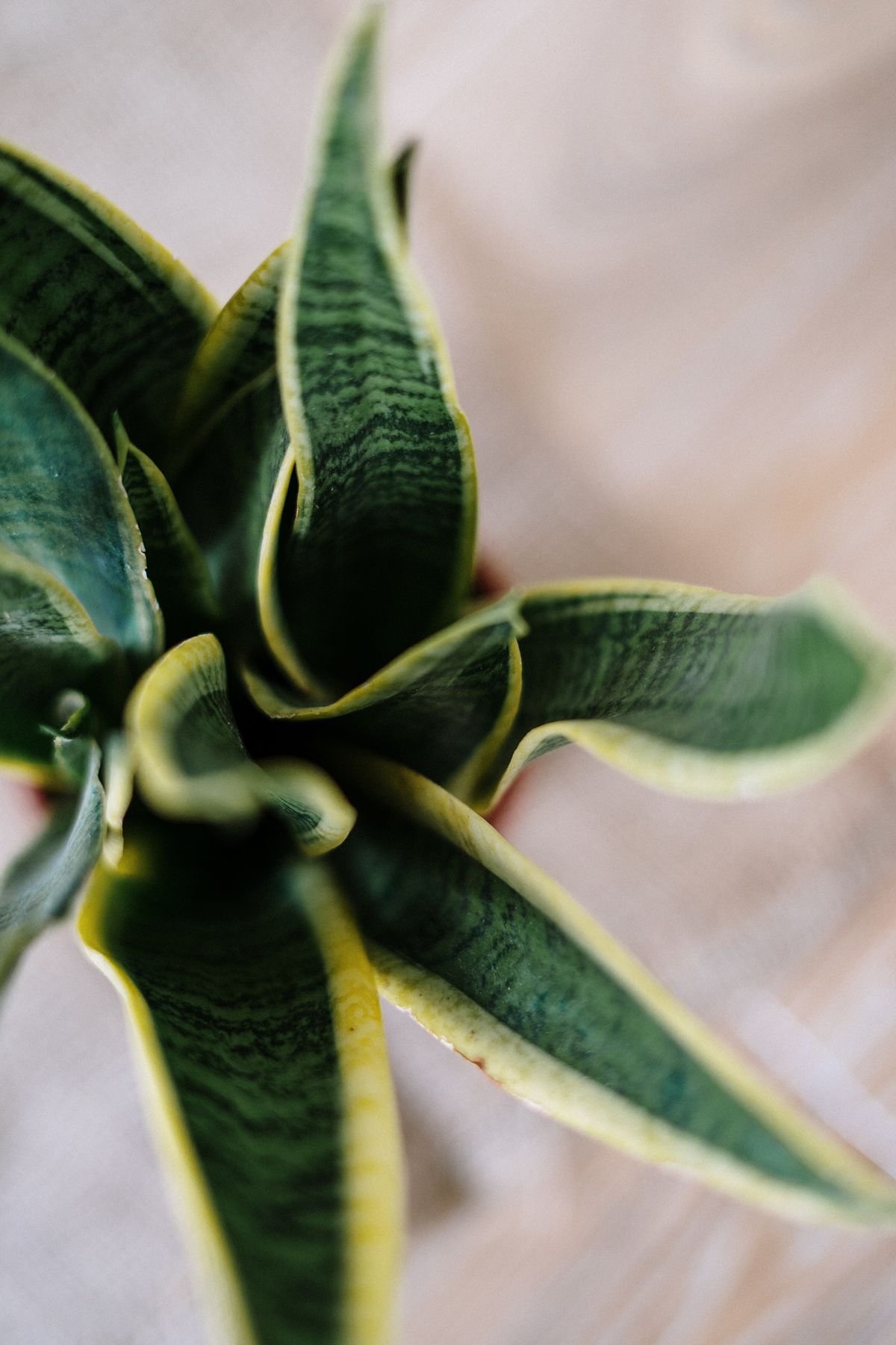
A snake plant leaf cross-section shows five distinct tissue layers: an outer waxy cuticle, an epidermis with stomata on both leaf surfaces, a ring of green photosynthetic chlorenchyma, a central colorless water-storage tissue called hydrenchyma, and structural fiber bundles reinforcing the whole structure. Each layer has one specific job, and together they explain nearly everything unusual about how this leaf looks, feels, and behaves.
Cut a snake plant leaf across its width and you are not looking at a simple green blade the way you would with most houseplants. You are looking at five genuinely different tissue types, nested inside each other like rings, each one solving a different problem the plant faces in dry, high-light conditions. (If you are trying to work out why a specific leaf looks or feels wrong, the Snake Plant Brown Spots guide covers diagnosing visible symptoms — this article covers the healthy structure those symptoms disrupt.) The Sansevieria Morphology guide covers how this leaf structure fits into the plant's overall growth habit.
The Snake Plant Leaf Cross Section, Layer by Layer
Working from the outside surface toward the center, a snake plant leaf is built from: a waterproof cuticle, an epidermis containing the stomata, a chlorophyll-rich chlorenchyma ring, a central hydrenchyma core for water storage, and fiber bundles woven through the whole structure for rigidity. Vascular bundles for transport run alongside the fibers rather than occupying their own separate layer. None of these tissues is unique to sansevieria on its own — what is unusual is the specific combination and arrangement, refined for a plant that needs to survive months without rain in direct sun.
Layer 1: The Waxy Cuticle
The outermost layer is a thick coating of wax sitting on top of the epidermis, giving the leaf its slightly polished feel and sheen. Its job is purely defensive: it dramatically slows water loss through the leaf surface itself, separate from whatever is happening at the stomata. The Sansevieria Adaptations guide covers this cuticle's role in the plant's overall drought strategy in full detail, including why dust buildup on this layer interferes with both water retention and light absorption.
Layer 2: The Epidermis and Its Stomata
Just beneath the cuticle sits the epidermis — a single outer layer of cells that includes the stomata, the small pores responsible for gas exchange. Here is a detail most general plant-anatomy explanations miss because it does not apply to typical broad, flat leaves: flat-leaved cultivars like Laurentii are amphistomatic, carrying stomata on both the upper and lower leaf surfaces rather than concentrating them on the underside only. This makes sense structurally — an upright leaf does not really have a shaded "underside" the way a horizontal leaf does, so both faces need their own gas-exchange capacity. Cylindrical species do not follow this same rule, and the Where Are Stomata Located on Snake Plant Leaves? guide covers that species-by-species difference in full.
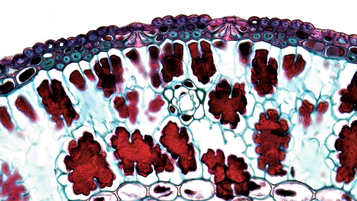
The stomata themselves are typically sunken into shallow grooves rather than sitting flush with the leaf surface, an arrangement that traps a small pocket of still, humid air right at the pore opening and further reduces water vapor loss — one more layer of the same xerophytic strategy running through this plant's entire structure. Their surrounding guard cells are also arranged irregularly rather than in the neat, predictable pattern seen in many broad-leaved plants, a configuration botanists classify as anomocytic — one more small structural signature of a leaf built for water conservation rather than rapid gas exchange. The timing of when these stomata actually open is a separate story covered in the CAM Photosynthesis in Sansevieria guide.
Layer 3: Chlorenchyma — the Photosynthetic Ring
Moving inward, the next distinct tissue is chlorenchyma: a band of chlorophyll-rich cells responsible for actually running photosynthesis. In a snake plant leaf, this photosynthetic tissue forms an outer ring rather than sitting in a single flat sheet the way it does in a typical broad leaf's palisade layer, wrapping around the leaf's outer portion just inside the epidermis on every side. This ring arrangement is a direct consequence of the leaf's shape — a flat leaf can afford a photosynthetic layer on just its two faces, but a thicker, more cylindrical or channel-shaped leaf needs that capacity distributed around its whole circumference to make efficient use of light hitting it from any angle.
Layer 4: Hydrenchyma — the Water-Storage Core
At the center of the leaf sits hydrenchyma, a colorless tissue with no photosynthetic role at all — its entire job is storing water. Research on Sansevieria leaf anatomy describes this water-storage tissue as forming "a highly branched 3-dimensional network of living cells" that fills the leaf's interior, distinct in structure and function from the chlorenchyma surrounding it. The How Do Snake Plant Leaves Store Water? guide covers exactly how this tissue holds and exchanges that water day to day, and the Why Do Snake Plant Leaves Become Wrinkled? guide covers what happens to it during drought — including the reinforced cell-wall bands that let it collapse in a controlled way rather than randomly.
Layer 5: The Fiber Bundles
This is the structural layer responsible for a snake plant leaf staying rigid and upright without any woody stem to lean on, and it is more complex than a single band of fiber running down the middle. Anatomical research on Sansevieria trifasciata documents two distinct fiber types working together: structural fiber bundles arranged in rows near the leaf margins on both the upper and lower surfaces, and separate ribbon or arc-shaped fibers scattered through the middle ground tissue rather than confined to the edges.
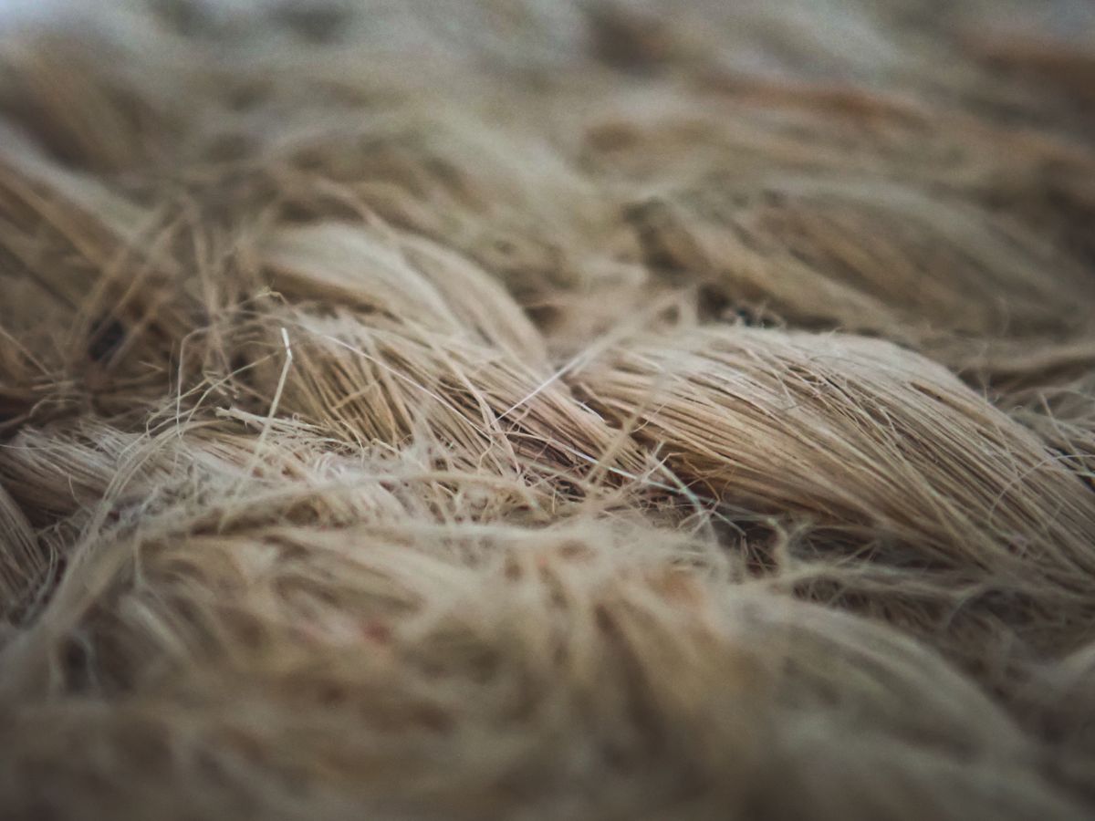
The same research found real, measurable differences between cultivars: bundles of roughly 150 cells in Laurentii's marginal fiber rows, compared to 29 or fewer cells in the compact Hahnii cultivar. That is not a minor technical detail — it is a plausible structural explanation for why Hahnii's compact rosette leaves feel noticeably softer and less rigid than a tall Laurentii leaf, independent of watering or light differences between the two. The Why Do Snake Plant Leaves Split or Crack? guide and the Why Do Snake Plant Leaves Lean or Fall Over? guide both cover what happens when this fiber network is pushed past its limits.
Running alongside these fiber bundles, rather than occupying a separate layer of their own, are the leaf's vascular bundles — the xylem and phloem responsible for moving water and sugars along the leaf's length. The close physical association between the vascular tissue and the fiber caps is not incidental: the same rigid bundles reinforcing the leaf mechanically also anchor and protect the more delicate transport tissue running beside them. The Snake Plant Vascular Bundles guide covers that vascular network — xylem, phloem, bundle sheath, and the monocot-wide parallel venation pattern behind it — in full.
How These Layers Work Together
None of these five layers does its job in isolation. The cuticle and sunken, amphistomatic stomata minimize water loss on both surfaces. The chlorenchyma ring captures light from whichever direction it arrives. The hydrenchyma core banks water reserves the whole leaf draws on during drought, its internal pressure directly contributing to how firm the leaf feels. The fiber bundles hold the entire structure upright, reinforced further by the turgor pressure of the hydrenchyma pressing outward from the inside — which is exactly why a severely underwatered leaf goes soft even though its fiber bundles remain physically undamaged. Losing water pressure does not break the scaffolding; it just removes the internal support that scaffolding was holding taut.
For the complete species profile these tissues belong to, the Sansevieria Plant Botanical Guide and the Sansevieria Trifasciata guide cover it in full.
Next time you trim a damaged leaf, look at the cut edge: the outer green ring is chlorenchyma, the pale, moist center is hydrenchyma, and the small tough strands running through both are the fiber bundles holding the whole thing together.
Care FAQ
What tissue layers make up a snake plant leaf cross section?
Five main layers from the outside in: a waxy cuticle, an epidermis with stomata on both leaf surfaces, an outer ring of chlorenchyma (photosynthetic tissue), a central hydrenchyma (water-storage tissue), and structural fiber bundles running through the mesophyll. Vascular bundles for water and nutrient transport run alongside the fiber bundles.
Does a snake plant leaf have stomata on both sides?
Yes. Unlike many broad, flat leaves that concentrate stomata on the underside only, sansevieria leaves are amphistomatic — they carry stomata on both surfaces, typically sunken into shallow grooves. This is a xerophytic adaptation that still allows gas exchange from either face of an upright, cylindrical, or channel-shaped leaf.
What is the difference between chlorenchyma and hydrenchyma in a snake plant leaf?
Chlorenchyma is the outer ring of green, chlorophyll-containing tissue that carries out photosynthesis. Hydrenchyma is the colorless water-storage tissue occupying the leaf's center, made of a branched network of cells whose main job is holding water in reserve rather than producing sugar. The two tissues sit in concentric layers, chlorenchyma outside, hydrenchyma inside.
Where are the fiber bundles located in a snake plant leaf?
In two distinct locations. Structural fiber bundles run in rows near the leaf margins, on both the upper and lower surfaces. A second type, ribbon or arc-shaped fibers, is scattered through the middle ground tissue rather than confined to the edges. Both types are made of thick-walled sclerenchyma cells and are the same fiber historically extracted for bowstring hemp.
Why do some snake plant cultivars have more fiber than others?
Fiber bundle count varies genuinely by cultivar, not just by individual plant health. Documented counts show considerably more cells per structural fiber bundle in Laurentii than in the compact Hahnii cultivar, consistent with Hahnii's generally softer, less rigid leaf structure compared to taller, stiffer varieties.
Does the cross-section structure explain why snake plant leaves are so stiff?
Largely yes. The combination of a rigid fiber-bundle network running the length of the leaf and turgor pressure inside the hydrenchyma is what keeps a snake plant leaf upright without a woody stem. Losing hydrenchyma turgor through underwatering softens the leaf noticeably, even though the fiber bundles themselves stay physically intact.